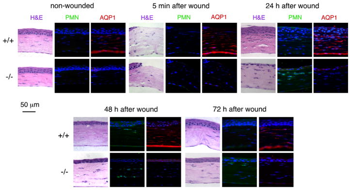
Fig. 3.
Healing of partial-thickness corneal stromal wounds in mice. H&E, neutrophils (green) and AQP1 (red) stained section from central mouse corneas. Nuclei stained with DAPI (blue). Micrographs shown for non-wounded corneas and at indicated times after wounding. (For interpretation of the references to colour in this figure legend, the reader is referred to the web version of this article.)

