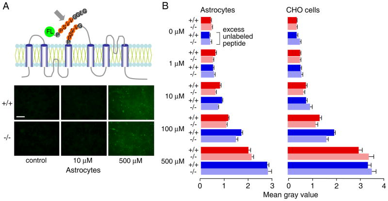
Figure 4. Lack of specific AQP4 binding of a putative AQP4 adhesion oligopeptide.
A. (top) Schematic of AQP4 membrane topology showing putative adhesion sequence (residues 139-142) in extracellular loop. The synthetic fluorescein-labeled 9-mer peptide (NH2-PPSVVGGLG-COOH) is shown. (bottom) Fluorescence micrographs of confluent astrocytes after incubation with indicated concentration of fluorescein-labeled 9-mer peptide for 30 min at room temperature, followed by brief fixation with 2% glutaraldehyde and washing. C. Summary of relative (background-subtracted) cell fluorescence for incubations done at indicated concentrations of the fluorescein-labeled 9-mer peptide, in the absence or presence of 50-fold excess unlabeled peptide (S.E., n=3–4). Differences not significant comparing AQP4 expressing vs. non-expressing glial and CHO cells. +/+: AQP4-expressing cells, −/− AQP4 null cells.

