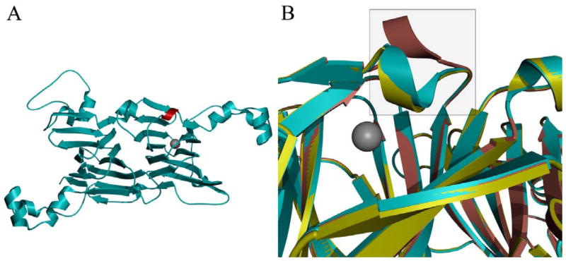
Fig. 2.
Ribbon representation of the protomer structure for the T165V OxDC variant (cyan) (A) with the SENS loop highlighted in red and the active site indicated by the location of the Mn(II) cofactor (grey sphere) and (B) overlaid with that of WT OxDC with the SENS loop (enclosed in square) in either the “open” (pink) or “closed” (yellow) conformation.

