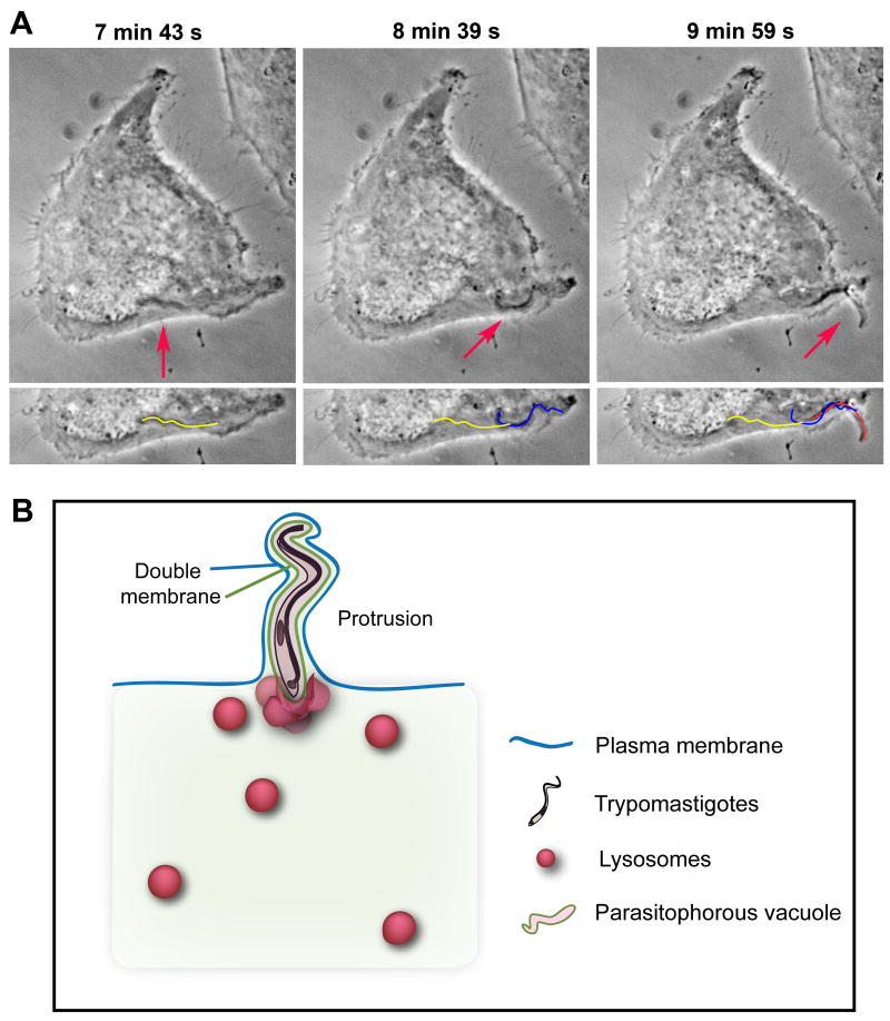
Figure 2. Internalized trypomastigotes protrude from host cells.
(A) Time Lapse live imaging (phase-contrast) of a trypomastigote moving inside a HeLa cell, and forming a plasma membrane protrusion (arrows). The yellow line represents the parasite's position at 7 min 43 s, the blue line at 8 min 39 s, and red line at 9 min 59 s (bottom panel). (B) Schematic model for protrusion event. Protruding trypomastigotes are surrounded by two distinct membranes: parasitophorous vacuole (green line), and plasma membrane (blue line). The parasites protrude in the direction of their flagellar movement (anterior end pointing outward) and stretch the overlying plasma membrane, while lysosomes progressively fuse with the vacuole and ultimately anchor trypomastigotes inside the cell.

