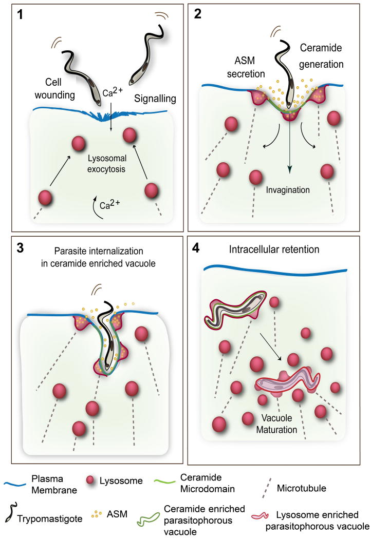
Figure 4. Model for T. cruzi cell invasion mediated by wounding and plasma membrane repair.
(1) Trypomastigotes wound mammalian cells, causing influx of extracellular Ca2+ and exocytosis of lysosomes. Signaling events also generate cytosolic Ca2+ transients. (2) Extracellular release of lysosomal ASM generates ceramide on the outer leaflet of the plasma membrane. (3) Ceramide-enriched plasma membrane microdomains facilitate trypomastigote internalization, while lysosomes continue to fuse with the nascent parasitophorous vacuoles, delivering ASM and generating additional ceramide. Lysosomal membrane anchoring to microtubules provides the force for pulling the parasites into the cells. (4) Recently internalized trypomastigotes residing in ceramide/Lamp1-enriched parasitophorous vacuoles fuse with additional lysosomes leading to parasite retention inside the cell.

