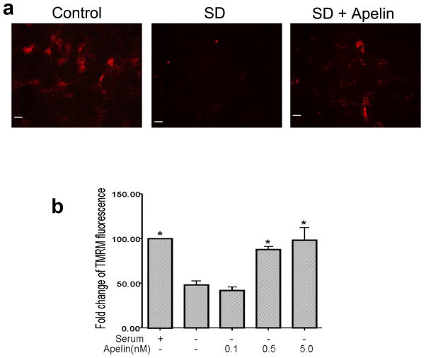
Figure 3. Effect of apelin-13 on the mitochondrial membrane potential.
The mitochondrial membrane potential was measured using TMRM fluorescence imaging in BMSCs. a. Control cells showed strong TMRM fluorescence indicative of normal membrane potential. After 12 hrs exposure to SD insult TMRM fluorescence decreased due to depolarization of the mitochondria. Apelin-13 (5 nM) added into the serum free media retained the mitochondrial membrane potential. b. Quantified data showing the protective effect of apelin-13 on the mitochondrial membrane potential. TMRM fluorescence decreased by 50% 12 hrs after SD while apelin-13 at 0.5 nM and 5 nM significantly reversed the loss of TMRM fluorescence. N=3 independent assays per group; Mean±SEM, * P<0.05 vs. SD group.

