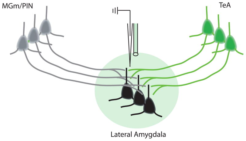
Figure 6. Optogenetic Control of Specific Afferent Inputs to the lateral nucleus of the amygdala (LA).
Infection of temporal association cortex (TeA) cells with an opsin expressing virus (an inhibitory opsin in this case) will produce opsin expression in TeA terminals in the LA. Green or yellow light (green sphere in figure) delivered through an in-dwelling fiber optic cable can then be used to inhibit the release of neurotransmitter specifically from TeA terminals while not affecting other inputs such as those from medial geniculate/posterior intralaminar thalamic nuclei (MGm/PIN, grey cells). When combined with in vivo neural recording in awake, behaving animals it would be possible to determine the contribution of TeA inputs to neural coding (for example of conditioned stimulus (CS) information) in LA neurons (black cells) and to fear behavior.

