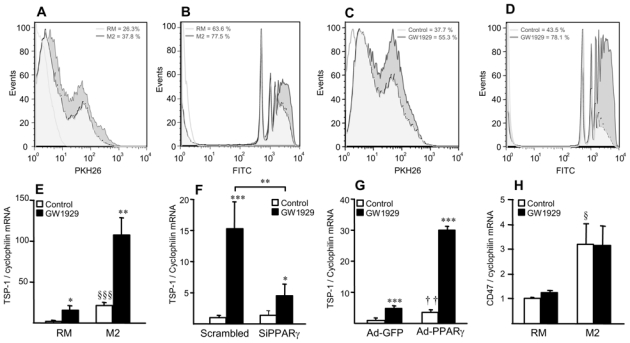
Figure 8. Alternative macrophage phagocytic activity is enhanced by PPARγ activation.

Panels A,B. Phagocytosis of apoptotic cells (A) and fluorescent beads (B) in RM and M2 macrophages.
Panels C,D. Phagocytosis of apoptotic cells (C) and fluorescent beads (D) in M2 macrophages in the absence or in the presence of GW1929 (600 nmol/L) for 24h. White histogram: isotype control.
Panels E–H. Q-PCR analysis of TSP-1 (E–G) and CD47 mRNA (H) in RM and M2 macrophages 24h-tretaed with GW1929 (600 nmol/L) (E,H), or in RM macrophages transfected with control or human PPARγ siRNA (F) or infected with Ad-GFP or Ad-PPARγ (G) and subsequently treated or not with GW1929 (600 nmol/L) during 24h. TSP-1 and CD47 mRNA levels were normalized to cyclophilin mRNA and expressed as means ± SD relative to control set at 1, from three independent experiments. Statistically significant differences are indicated (t-test; M2 vs RM, §p<0.05, §§§p<0.001; Ad-PPARγ vs Ad-GFP, ††p<0.01; GW1929-treated vs control, *p<0.05, **p<0.01, ***p<0.001).
