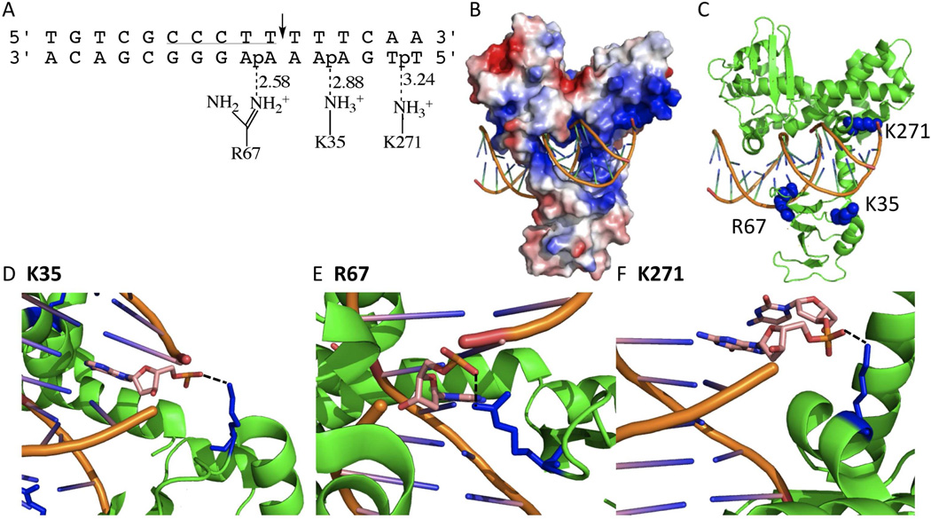
Figure 1.
Variola topoisomerase-DNA interactions (PDB ID 3IGC). (A) Schematic of K35, R67, and K271 interactions with duplex DNA. Dashed lines indicate hydrogen bonds with the distance in angstroms. The vTopo DNA recognition sequence is underlined, and cleavage occurs at the phosphate indicated by the arrow. (B) Global electrostatic map of vTopo complexed with DNA, red and blue indicates negative and postive electrostatic potential, respectively. DNA is shown in stick mode (orange). (C) Same structure from (B) with the mutated residues shown as blue spheres and the protein and DNA shown in green and orange. (C) Interaction of Lys35 with the +3 phosphate on the uncleaved strand, (D) Interaction of Ag67 with the −1 phosphate on the uncleaved strand, (C) interaction of Lys271 with the +6 phosphate on the uncleaved strand. Panels (B–F) were produced using PyMol (17).

