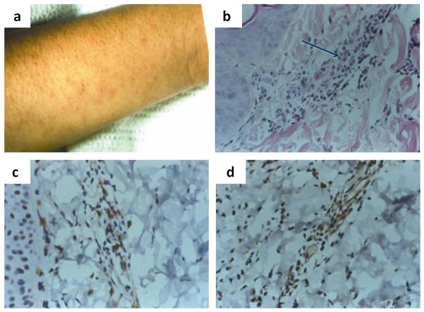
Figure 3.
The measles virus rash (a) is indicative of the immune response and results from the infiltration of leukocytes (b), including CD4+ (c) and CD8+ (d) T lymphocytes into sites of virus replication in the skin. Histological examination of a biopsy of a measles skin rash lesion shows (a) an accumulation of mononuclear cells (arrow) that have infiltrated an area of infected epithelial cells (hematoxylin and eosin stain). Immunoperoxidase staining (brown) of the biopsy for CD4+ (c) and CD8+ (d) T cells shows that many of the infiltrating mononuclear cells are T lymphocytes (Polack et al, 1999).

