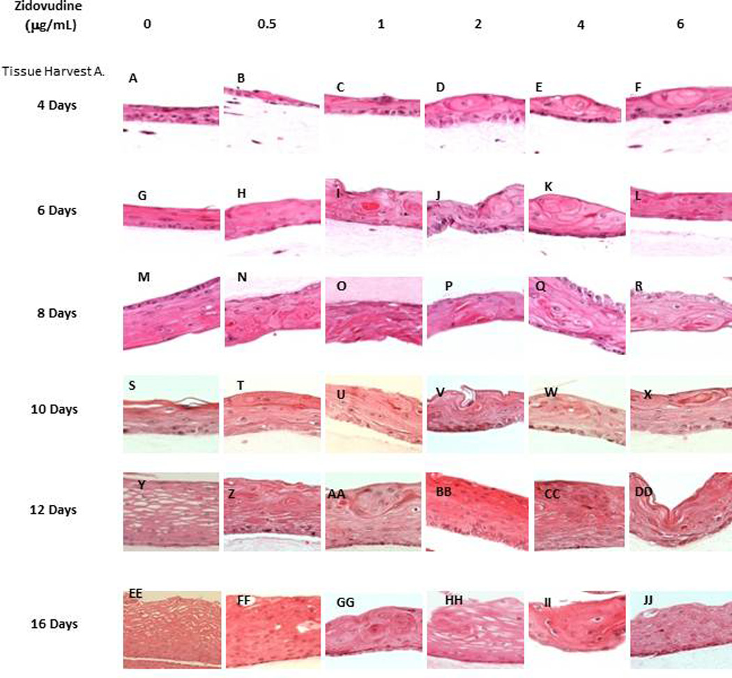
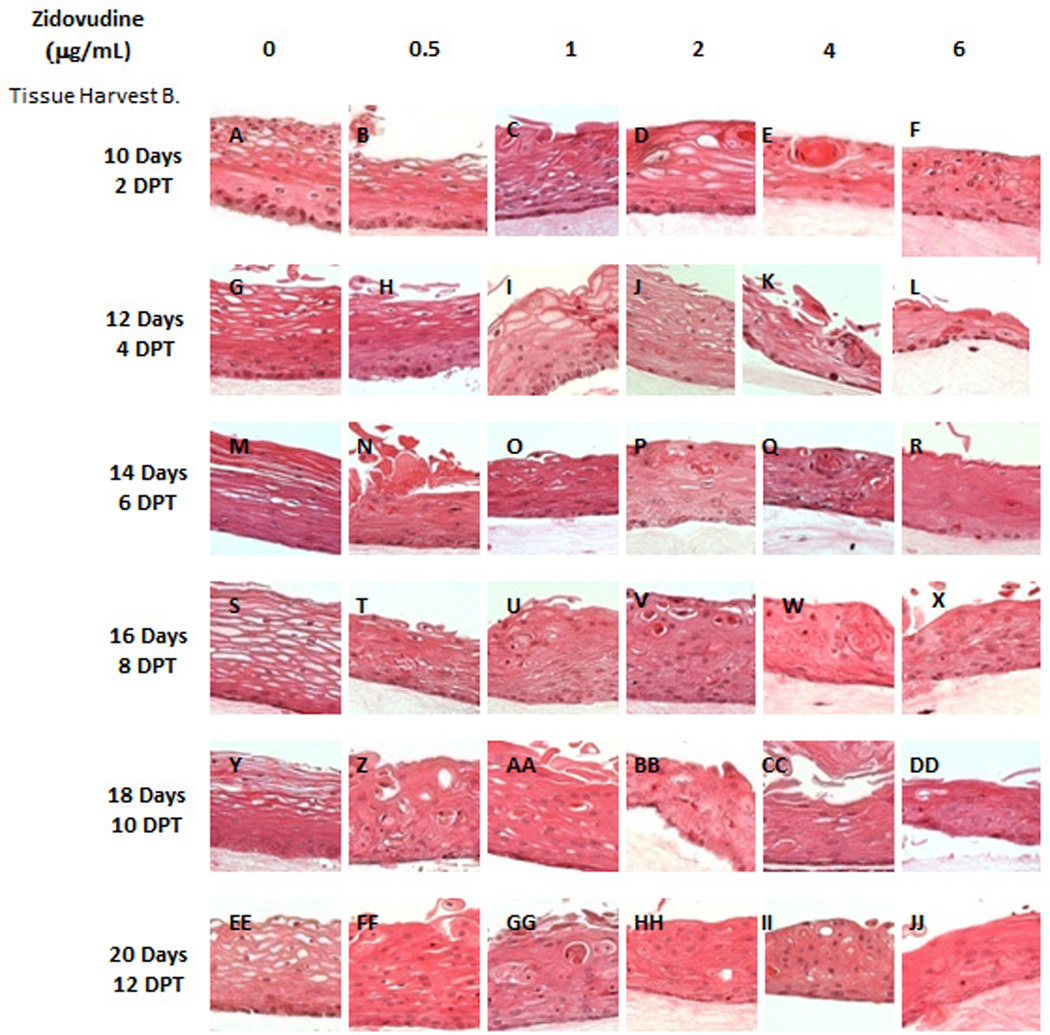
Figure 2. Effect of Zidovudine on gingival epithelium morphology and stratification.


Primary gingival keratinocytes were grown in organotypic (raft) cultures and treated with different concentrations of Zidovudine, beginning either at day 0 (Tissue Harvest A) or at day 8 (Tissue Harvest B). (Panels A, G, M, S, Y and EE) untreated rafts; (Panels B, H, N, T, Z, and FF) rafts treated with 0.5 µg/ml Zidovudine; (Panels C, I, O, U, AA, and GG) rafts treated with 1 µg/ml Zidovudine; (Panels D, J, P, V, BB and HH) rafts treated with 2 µg/ml, the cMax of Zidovudine; (Panels E, K, Q, W, CC, and II) rafts treated with 4 µg/ml Zidovudine; (Panels F, L, R, X, DD, and JJ) rafts treated with 6 µg/ml Zidovudine. Rafts were harvested at different points and stained with hematoxylin and eosin. DPT denotes days post treatment (Tissue Harvest B). Images are at 20 X original magnification.
