
Figure 8. MC3R agonist increases the number of pERK-IR neurons in the DMH.
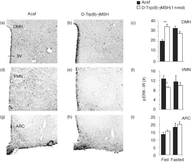
pERK-IR neurons in the DMH (a and b), VMN (d and e) and ARC (g and h) in control B6 mice treated with Acsf (a, d and g) or 1 nmol d-Trp(8)-γMSH (b, e and h) fed ad libitum or after an overnight fast. The representative photomicrographs were taken using sections collected from mice treated in fed conditions. Treatment with d-Trp(8)-γMSH significantly increased the number of pERK-IR neurons in the DMH in fed (representative photomicrograph at bregma level: −1.82 mm), but not fasting conditions (c). In the VMN, d-Trp(8)-γMSH significantly reduced the number of pERK neurons (f) in fed, but not fasting conditions (representative photomicrograph at bregma level: −1.58 mm). There was no effect of treatment in the ARC (i) (representative photomicrograph at bregma level: −1.58 mm). *P < 0.05 vs. Acsf.**