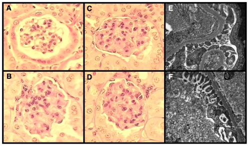
Figure 4. Glomerular histopathology of diabetic Akita mice.
Photomicrographs of mouse kidneys were taken using tissue samples prepared from mice at 30-weeks of age. (A) A representative picture from a control mouse (score of 0). (B–D) Moderate mesangial expansion in glomeruli from Akita mice (score of 2). (E) A representative picture of glomerular ultrastructure from a control mouse. (F) FP effacement was not detected by electron microscopy in diabetic Akita mice at 30-weeks of age. Light microscopic sections were stained with Periodic acid-Schiff (PAS) and the magnification was 400×.

