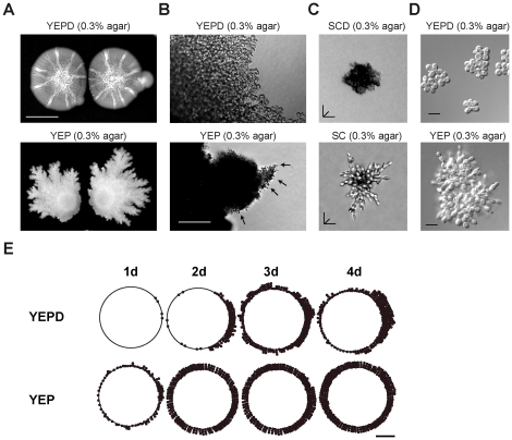
Figure 1. S. cerevisiae forms filamentous mats.
A) Wild-type cells (PC313) were spotted 2 cm apart onto 0.3% agar media that contained (YEPD; top panel) or lacked (YEP; bottom panel) glucose. The YEPD plate was incubated for 4 days and photographed; the YEP plate for 15 days. Bar = 1 cm. B) Microscopic examination of perimeters of mats in 1A. Bar = 100 microns. C) The origin of filamentous mats. Wild type (PC538) cells were examined on synthetic medium either containing 2% glucose (SCD) or lacking glucose (SC) in 0.3% agar medium for 24 h at 30°C. A compiled Z-stack rendering of typical microcolonies are shown. Bar = 20 microns. D) Same strains in 1C were examined on rich medium either containing 2% glucose (YEPD) or lacking glucose (YEP) in 0.3% agar. A representative microscopic image is shown. Bar = 10 microns. E) Vegetative mats mature into filamentous mats over time as nutrients become limiting. Two mats of wild type (PC313) strain were spotted bilaterally (1.5 cm apart) on YEPD and YEP media (+0.2% galactose) containing 0.3% agar media. The number of filaments occurring along the circumference of mats was scored on a scale of 1, 2, or 3 dots at 20× magnification corresponding to 3, 6, or 9 filaments or greater, respectively. Dots were plotted on a circle representing the outline of one of the mats with right hemispheres corresponding to the side of the mat facing a second mat. Asymmetric filamentation observed in the right hemisphere of 2d, Glu can possibly result from nutritional stress compounded by nutrient depletion from adjacent mats. Filamentation was monitored and plotted after growth for 1, 2, 3, and 4 days. Quantitation of pseudohyphae was complicated at longer time points when biofilms began to variegate [60]. Bar = 1 cm.

