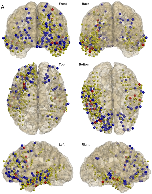
Figure 6. Spatial distribution of phase-modulated gamma patterns.
Locations of all intracranial contacts associated with IN-phase (blue balls) and ANTI-phase (red balls) patterns for 11 subjects for which Talairach coordinates could be successfully estimated are superimposed on a cortical reconstruction of one subject. Yellow balls correspond to the locations of all analyzed contacts for the corresponding 11 subjects (418). From left to right and from top to bottom, views are presented from the following planes: frontal-coronal, back-coronal, top-axial, bottom-axial, left-sagital and right-sagital.

