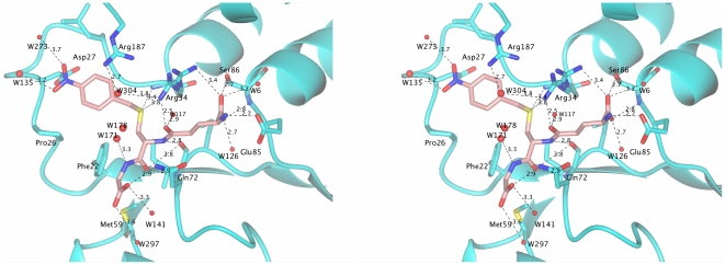
Figure 4. Close-up stereo view of the active site. Hydrogen-bonds (<4.0 Å) between Nb-GSH and the enzyme are shown as dashed lines.
W304 and W117 from the proposed electron-sharing network are depicted. The orientation of Nb-GSH is the same as in Figure 3C. The figure was produced using the CCP4 molecular graphics program [56].

