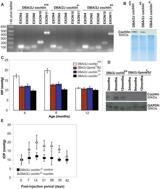
Figure 2. Down-regulation and/or disruption of cochlin expression in mice TM is commensurate with reduction in mean IOP.
(A) Agarose gel (1.2%) showing the absence of PCR products for exons 8, 10 and 11 in the DBA/2J cochlin−/− mice. These exons are present in DBA/2J cochlin+/+ and cochlin+/− mice. DNA was isolated from mice tail and subjected to PCR amplification for the indicated exons. (B) Western blot analysis of TM extract of DBA/2J cochlin+/+, DBA/2J cochlin+/− and DBA/2J cochlin−/− mice showing the complete absence of cochlin in DBA/2J cochlin−/− mice. Coomassie blue stained gel shows equal loading of protein in all the lanes. (C) Intraocular pressure (IOP in mmHg) in different mice strains at indicated ages (n = 35–45 for each strain at each age group). The DBA/2J cochlin+/+ mice show a higher mean IOP as compared to DBA/2J-Gpnmb+/SjJ, DBA/2J cochlin+/− and DBA/2J cochlin−/− mice at 6 months and at 8 months of age. The difference is most marked at 8 months of age. By 12 months, the mean IOP is similar for the different strains. (D) Western blot analysis of the TM protein extract from the DBA/2J cochlin+/+ and DBA/2J-Gpnmb+/SjJ at the indicated ages, probed for cochlin. GAPDH has been shown as a loading control. (E) Generation 10 (Gen 10) DBA/2J cochlin−/− at six months of age were injected with a lentiviral vector bearing the COCH-GFP transgene (n = 20) or GFP alone (n = 20) in the anterior chamber. The mice were followed and IOP recorded at the indicated time periods. Error bars (C and E) depict ± standard deviation.

