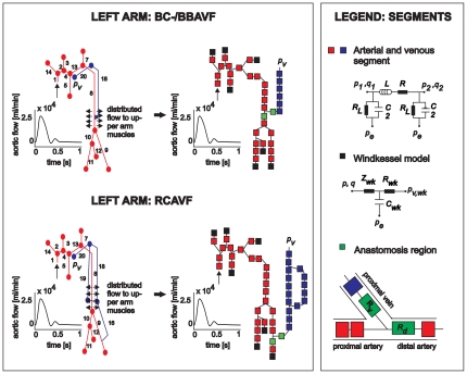
Figure 1. Left arm vasculature divided into arterial, venous and anastomosis segments (middle).
These segments locally describe the relation between pressure p and flow q via a lumped parameter approach (right), and consists of a resistor R (viscous resistance to blood flow), a resistor RL (viscous resistance of blood flow to small side-branches), an inductor L (blood inertia) and a capacitor C (vascular compliance). The anastomosis is modeled with two nonlinear resistors Rv and Rd. The windkessels consist of two resistors, Zwk and Rwk (together the peripheral resistance) and a capacitor Cwk (peripheral compliance). This figure is adapted from Huberts et al.

