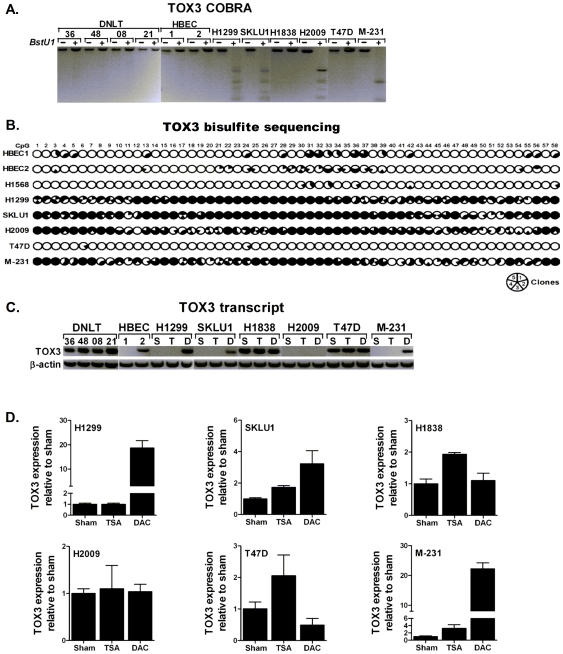
Figure 4. Methylation and expression of TOX3 in lung and breast cancer.
(A) COBRA and (B) bisulfite sequencing assays were used to assess the methylation status of TOX3 and the results are summarized as described for figure 1. (C) Expression of TOX3 and beta-actin in DNLT, HBEC, and various lung and breast cancer cell lines. TOX3 was silenced in vehicle-treated (S) lung cancer (H1199, SKLU1, and H2009) and breast cancer (MDA-MB-231, abbreviated as M-231) cell lines with methylated promoter CpG island. Treatment with 5-Aza-2′-deoxycytidne (D) but not trichostatin A (T) restored TOX3 expression. (D) Quantitative analysis of TOX3 in lung and breast cancer cell lines treated with Vehicle (Sham), TSA, and DAC using a TaqMan assay that uses primer sets distinct from the primers used for gel-based assays confirmed results shown in figure 3C.

