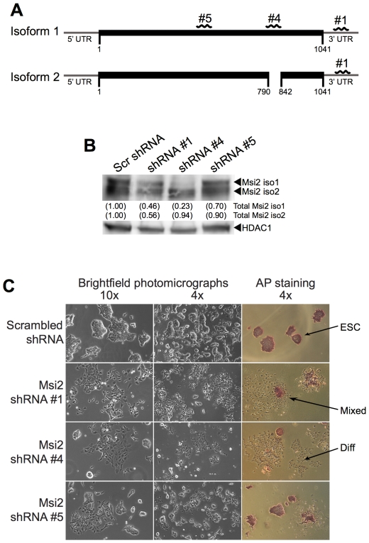
Figure 2. Knockdown of Msi2 results in the differentiation of ESC.
(A) Regions of Msi2 mRNA targeted by shRNA #1, shRNA #4, and shRNA #5. (B) The D3 ESC were infected with lentiviruses that express scrambled (Scr) shRNA, shRNA #1, shRNA #4, or shRNA #5 sequences. Two days after infection, the cells were subjected to puromycin selection for 24 hours. After selection, the cells were subcultured and grown for an additional 24 hours before nuclear extracts were harvested for western blot analysis. HDAC1 was used as the loading control for quantification. (C) Bright field photomicrographs of cells subcultured at 5,000 cells per cm2 were taken 7 days post-infection with each shRNA (left columns). Cells were stained with alkaline phosphatase (right column) 10 days post-infection after being subcultured at 200 cells per cm2. Arrows point to colonies that exhibit a morphology characteristic of ESC (ESC), a morphology consisting of ESC and differentiated cells (Mixed), or a morphology characteristic of differentiated cells (Diff).

