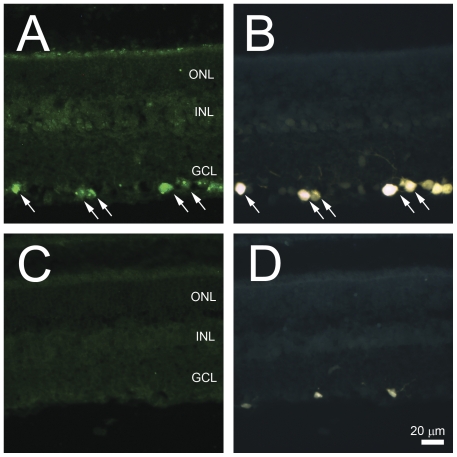
Figure 2. Immunohistochemical localization of Nell2 in control and ONT retinas.
A and B. Co-localization of Nell2 expression with FG-labeled RGCs. The distribution of Nell2 protein in the retina was similar to its mRNA expression. The most abundant expression of these proteins was observed in the GCL. Furthermore, all Nell2-expressing cells in the GCL were colocalized with retrogradely-labeled RGCs. C and D. Loss of Nell2 immunohistochemical staining correlates with the loss of RGCs 2 weeks after ONT. Analysis of Nell2 protein expression 2 weeks after ONT showed no Nell2-positive cells in the retina. Very few FG-positive microglial cells are present.

