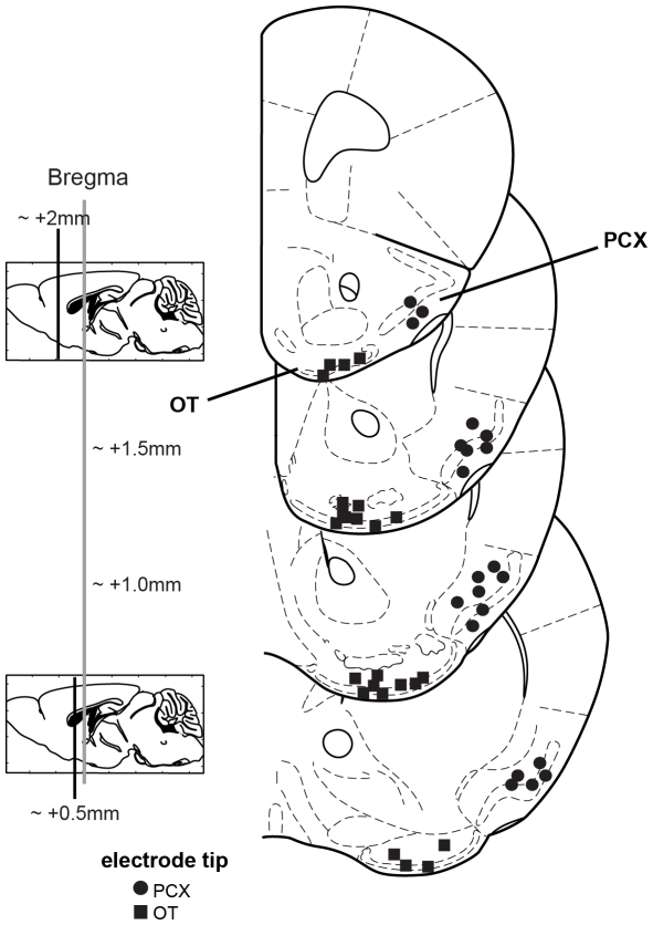
Figure 1. Electrode tip locations verifying extent of OT and PCX recording sites.
Coronal stereotaxic panels showing the approximate location of electrode tips from records used for analysis. Coronal sections span from 2.0 – 0.5 mm anterior of bregma, in 0.5 mm intervals. Panels adapted from [61].

