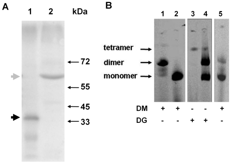
Figure 3. γ-25-HcRed fusion proteins crosslink mtATPase complexes.
A. Mitochondrial lysates were subjected to SDS-PAGE and blots probed with antisera against mtATPase subunit γ. Lane 1, YRD15 control; and lane 2, γ-25- HcRed The position of molecular weight markers are indicated at right. The positions of the γ fusion protein and endogenous subunit γ are indicated at left by gray and black arrowheads, respectively. B. Mitochondria isolated from cells were solubilised with the addition of dodecyl β-maltoside (DM) or digitonin (DG) as indicated, and lysates subjected to clear native-PAGE. Gels were imaged for fluorescence: γ-25-HcRed (lanes 1 and 3); γ-25-mRFP (lanes 2 and 4), and γ-25-HcRed: mtHcRed (lane 5). The positions of monomer, dimer and tetramer mtATPase complexes are indicated at left.

