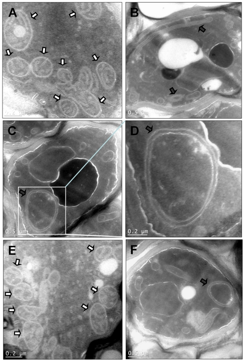
Figure 4. Mitochondria in yeast cells expressing γ-25-HcRed fusion proteins lack cristae.
Transmission electron microscopy was performed on cell sections. A, YRD15; B - D, γ-25-HcRed; E, γ-25-mRFP, and F, γ-25-DsRed. Boxed area in C is shown magnified in D. Mitochondria and abnormal mitochondria are highlighted by white and black arrows respectively. Scale bars are shown.

