
Figure 8.
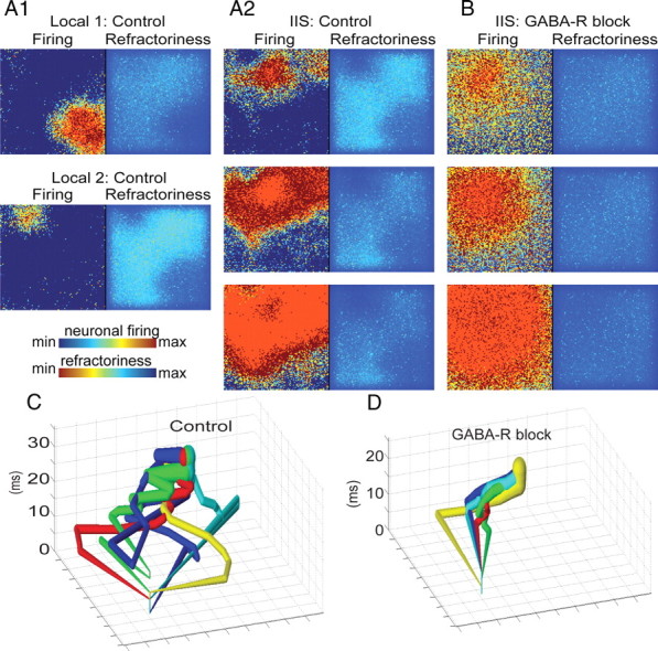
GABAergic networks determine the IIS pathways via the creation of refractory pockets. A1, A2, When inhibition was intact in our model of epileptiform activity (control), spontaneous IIS events were preceded by a series of local events (A1) in which spatially restricted, transiently synchronous activity (red regions, left) was quenched by local activation of GABAergic synapses, resulting in pockets of refractoriness due to activity-dependent, short-term depression of glutamatergic synapses (dark blue regions, right). Upon initiation of an IIS (A2, top), activity propagates (center and bottom) evenly through nonrefractory areas (light blue and green regions, right), but poorly through refractory regions. B, When GABAergic synapses were blocked locally, synchronized, nonpropagating events no longer occurred; rather, the first synchronization spread evenly through the entire neural network. C, D, We applied the spatial averaging technique described in Figure 1 to the neuronal firing output of our computational model to track the propagation of IISs initiating from a single spatial location before and after GABA-R blockade. In agreement with our in vitro data, GABA blockade sharply reduced the spatial variance of the propagation of synchronous activity through the network (average distance between each trajectory control vs GABA-R block; t test, p < 0.01).