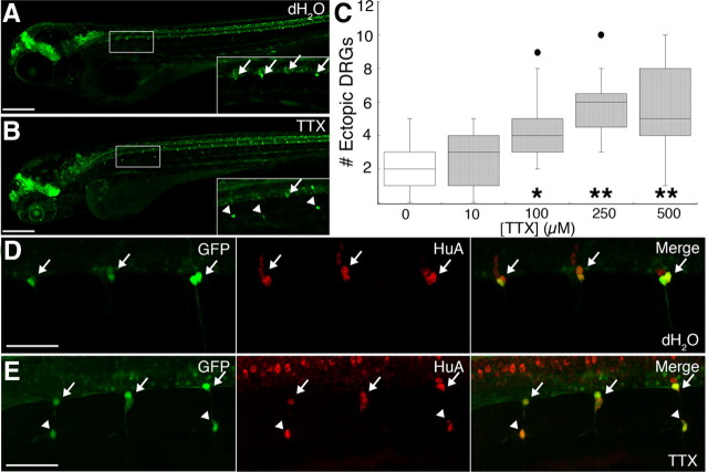
Figure 1.
Maintenance of DRG position requires sodium current. A, B, Whereas DRG neurons (arrows) (A) are regularly arranged along the ventral boundary of the spinal cord in 4 dpf Tg(-3.4neurog1:GFP) dH2O-injected embryos, DRG cells (B) are absent from this location in several segments and GFP-expressing cells are instead located in ectopic ventral positions (arrowheads) in sibling embryos injected with 250 μm TTX. Insets correspond to the boxed regions in A and B. C, The median number of ectopic DRG neurons per embryo at 4 dpf increases in a dose-dependent manner with TTX concentration (*p < 0.05 vs 100; **p < 0.001 vs 250, and 500 μm TTX, nonparametric Kruskal–Wallis test; sample sizes ranged between 15 and 22 embryos). The graph presents the inner quartiles as a box with an internal line indicating the median; whiskers extend to the 5th and 95th percentiles; filled circles indicate outliers. D, E, In 4 dpf Tg(-3.4neurog1:GFP) embryos, both normally positioned as well as ectopic GFP-expressing DRG cells (arrows and arrowheads, respectively) express HuA (red), a marker of neuronal differentiation. Scale bars; A, B, 200 μm; D, E, 50 μm.

