Figure 6.
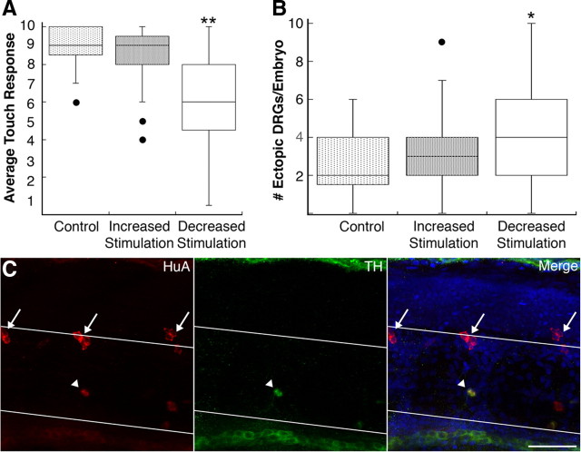
Decreased tactile stimulation leads to ventral migration and transdifferentiation of DRG neurons. A, The 4 dpf embryos raised under conditions that decrease tactile stimulation show a reduced touch response compared with embryos raised under control or increased stimulation conditions (**p < 0.001 vs control and increased stimulation, Kruskal-Wallis; sample size ranged between 10 and 65 embryos). B, Decreased tactile stimulation increases the number of ectopic DRG neurons at 4 dpf (*p = 0.02 vs control, Mann–Whitney U test; sample size ranged between 20 and 50 embryos). C, At 11 dpf, a subset of migratory DRG neurons (arrowhead) expresses the sympathetic marker TH (green). HuA immunoreactivity is shown in red; white lines demarcate the dorsal and ventral boundaries of the notochord; arrows indicate normally positioned DRG neurons. The fraction of migratory DRG neurons expressing TH does not differ significantly between control (9/35; 14 embryos), increased stimulation (17/58; 16 embryos), and sensory-deprived embryos (16/47; 13 embryos). Scale bar, 40 μm.

