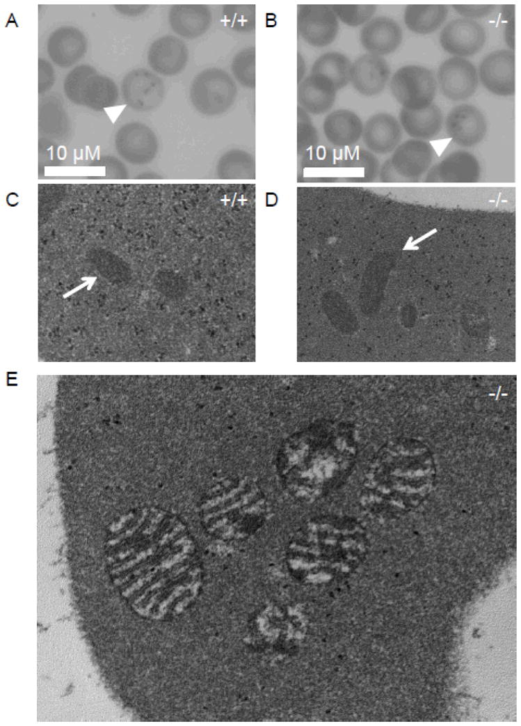
Figure 3. Microscopic evaluation of Ftmt-deficient erythrocytes.

(A,B) Light micrographs of Prussian Blue-stained blood smears from wild-type (+/+) (A) and Ftmt-deficient (−/−) (B) mice; siderocytes are indicated by arrowheads; 40x magnification. (C,D) Electron micrographs of reticulocytes from wild-type (+/+) (C) and Ftmt-deficient mice (−/−) (D); mitochondria are indicated by arrows; 40,000x original magnification. The wild-type and mutant mitochondria are indistinguishable. (E) High-power electron micrograph of a siderocyte from a Ftmt-deficient mouse; 80,000x original magnification. Note the slight enhancement of the mitochondrial membranes and amorphous electron dense deposits detailed in (E), which are similar to previous descriptions of murine siderocytes by electron microscopy(20). All samples were obtained from mice on pyridoxine-deficient diets. All blood samples were stained with acid ferrocyanide prior to processing for electron microscopy.
