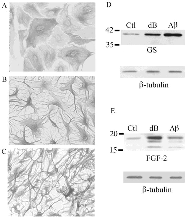
Figure 1. Reactivity observed in primary astrocytes treated with dB-cAMP or Aβ.
(A) Vehicle-treated control (Ctl) neonatal astrocyte cultures show polygonal morphology. In contrast, stellate morphology is observed in cultures treated with (B) 1 mM dBcAMP (dB) or (C) 25 μM Aβ25–35. Representative western blots demonstrate increased expression of (D) GS and (E) FGF-2 in cultures treated for 48 h with dBcAMP (dB) or Aβ relative to vehicle-treated controls (Ctl).

