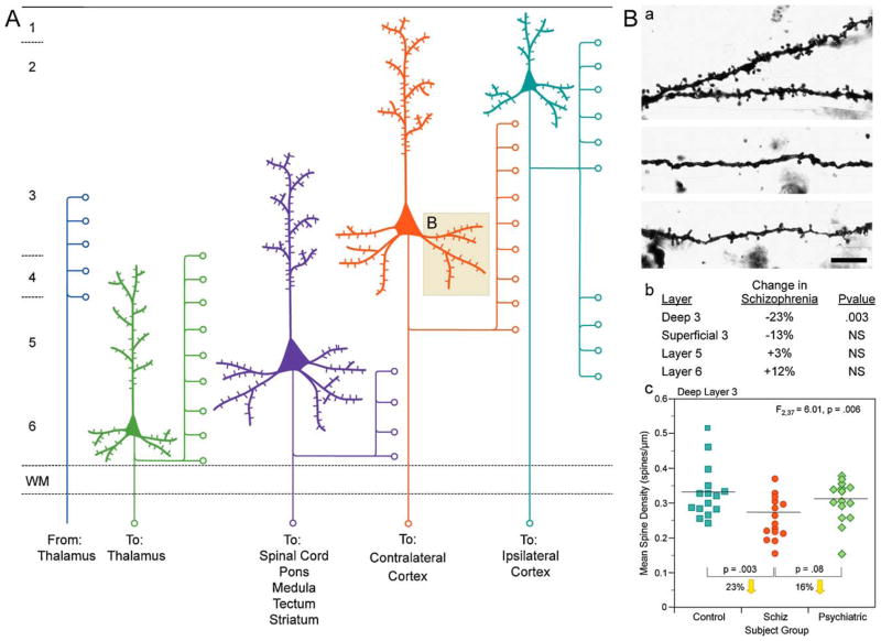
Fig. 1.
(A) Schematic representation of the main afferent and efferent pyramidal connections to and from specific cortical lamina. (B) Reduction in pyramidal neuron dendritic spines in deep layer 3 of the DLPFC in schizophrenia. (a) Golgi-impregnated basilar dendrites and spines on deep layer 3 pyramidal neurons from a normal comparison (top) and two subjects with schizophrenia (bottom). Note the reduced density of spines in the subjects with schizophrenia in these extreme examples. (b) Laminar specificity of the spine density differences in the subjects with schizophrenia relative to normal control subjects. (c) Scatter plot demonstrating the lower density of spines on the basilar dendrites of deep layer 3 pyramidal neurons in the DLPFC of subjects with schizophrenia relative to both normal and psychiatrically ill comparison subjects.
Adapted from Lewis and Gonzalez-Burgos (2008).

