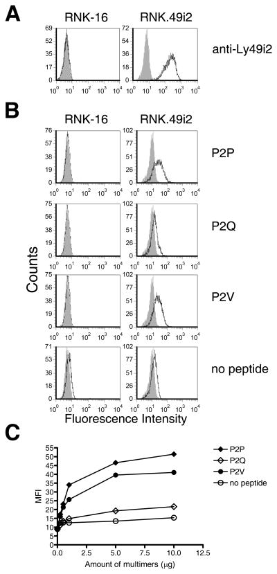
FIGURE 4. RT1-A1c bound with P2 anchor residues Pro or Val, but not Gln, bind to Ly49i2 expressing cells.
A. RNK-16 and RNK-16 cells stably transfected with Ly49i2 (RNK.49i2) were stained with STOK2 (anti-Ly49i2) Ab (open histograms) or isotype control (grey histograms) B. RNK-16 and RNK.49i2 cells were stained with PE-labeled RT1-A1c multimers bearing the peptides P2P, P2Q, or P2V, or no peptide (open histograms) or Extravidin-PE alone (grey histograms). C. Increasing amounts of PE-labeled RT1-A1c multimers were used to stain RNK.49i2 cells. The extent of staining was determined by FACS analysis and plotted as MFI vs. amount of multimers. RT1-A1c was refolded with P2P, P2Q, and P2V and purified three separate times with similar cell-staining results.

