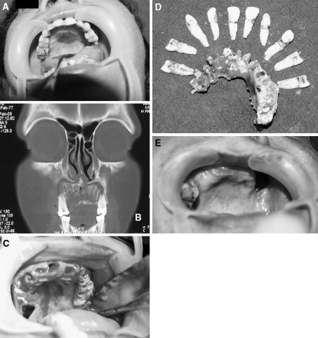
Fig. 2.
a Patient 2 Intra Oral picture of a patient with Maxillary Osteomyelitis. b Coronal section of the CT scan showing Osteomyelitic Maxilla. c Intra operative view after extraction of teeth showing the necrosed bone. d Sequestrum with the extracted teeth. e Intra Oral view showing well healed Maxillary Osteomyelitis with Oro Antral Fistula 1.5 year post operative

