
Figure 2.
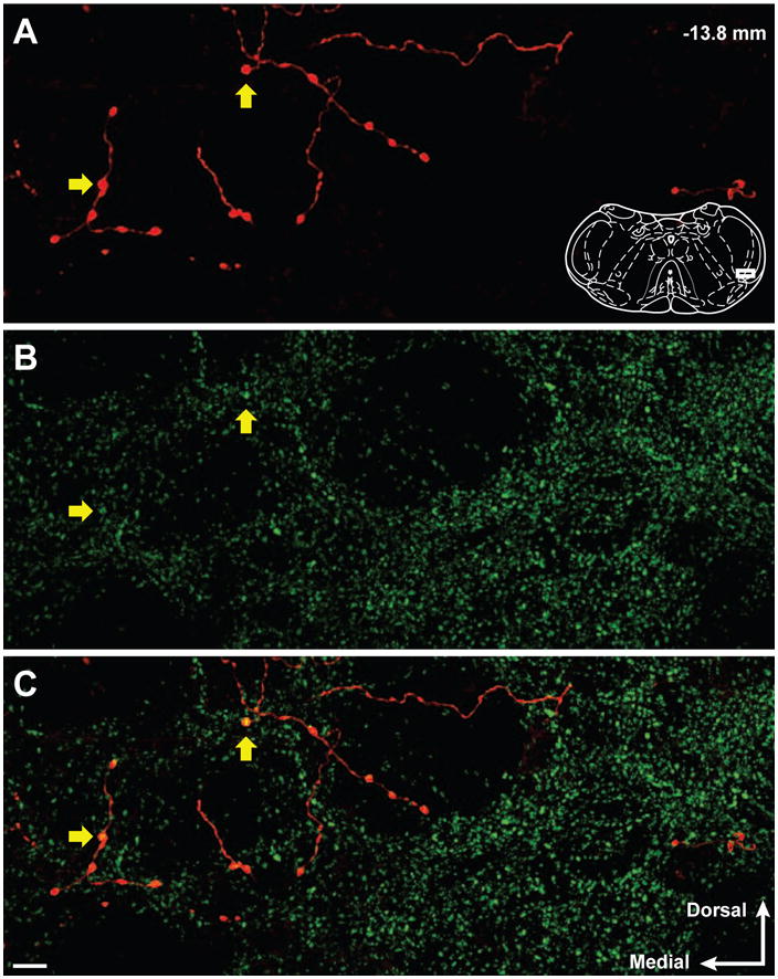
Confocal micrographs show BDA-labeled reticulomedullary varicosities and GAD67 immunoreactivity in the trigeminal dorsal horn. (A) Reticulomedullary projections display axonal labeling with distinct varicosities. (B) GAD67 immunoreactivity in the trigeminal dorsal horn is abundant and predominantly punctate. (C) Overlay of panels A and B illustrate several BDA-labeled reticulospinal varicosities that contain GAD67 immunoreactivity (Yellow arrows). The representative diagram of the trigeminal brainstem in panel A is modified from the digital rat atlas of Paxinos and Watson (Paxinos and Watson, 1998) and is reproduced here with permission from the publisher. The white rectangle on the diagram in panel A represents the location of the micrographs. The number in the upper right of panel A represents the distance from bregma. The white arrows in the lower right of panel C represent the medial and dorsal orientation of the micrographs. Micrographs are 14 consecutive and overlapping optical sections composing a 6.5 um thick Z stack. Scale bar = 10 um.