
Figure 3.
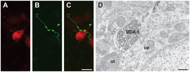
Reticulomedullary and reticulospinal terminals were observed contacting neurons with confocal (A-C) and electron (D) microscopy. (A-C) Confocal micrographs (10 consecutive and overlapping optical sections composing a 4.5 um thick Z stack) illustrate (A) a NeuN-ir neuron and (B) BDA-labeled reticulomedullary varicosities in the trigeminal dorsal horn. (C) The overlay demonstrates appositions between two BDA-labeled reticulomedullary varicosities and the NeuN-ir cell. Scale bar = 10 um for panels A - C. (D) Electron micrograph illustrating a BDA-labeled terminal (BDA-t) forming an apposition with an unlabeled perikarya (up). An unlabeled terminal (ut) is also seen forming an apposition with the cell. Scale bar = 500 nm.