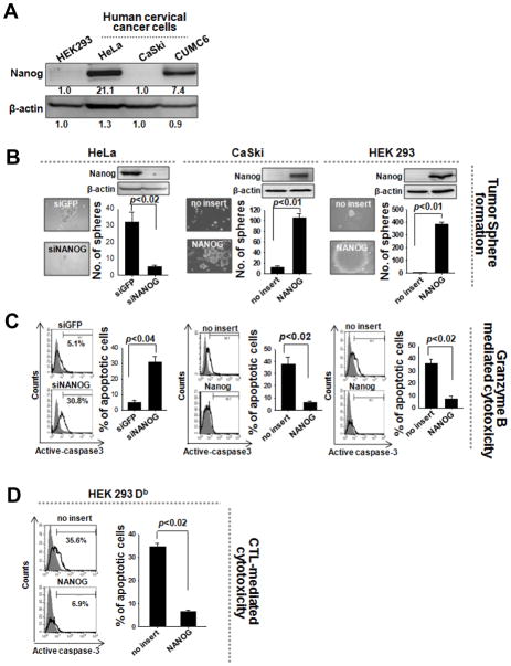
Figure 7. Nanog-mediated control of stemness and immune escape in human cancer.
(A) Western blot was performed to characterize Nanog expression in human cervical cancer cells. (B) CUMC6 or HeLa cells with high levels of Nanog were treated with siGFP or siNanog. CaSki or HEK293 cells with low levels of Nanog were transduced with empty vector or NANOG. The sphere-forming capacity of these cells was analyzed in suspension culture. (C) Recombinant GrB was delivered into the cells. The frequency of apoptotic (active caspase-3+) cells was measured by flow cytometry. (D) Empty vector- or Nanog-transduced HEK293Db cells were pulsed with E7 peptide and then mixed with E7-specific CTLs at a 1:1 ratio. The percentage of apoptotic cells was quantified by flow cytometry. Data are representative of at least three independent experiments.

