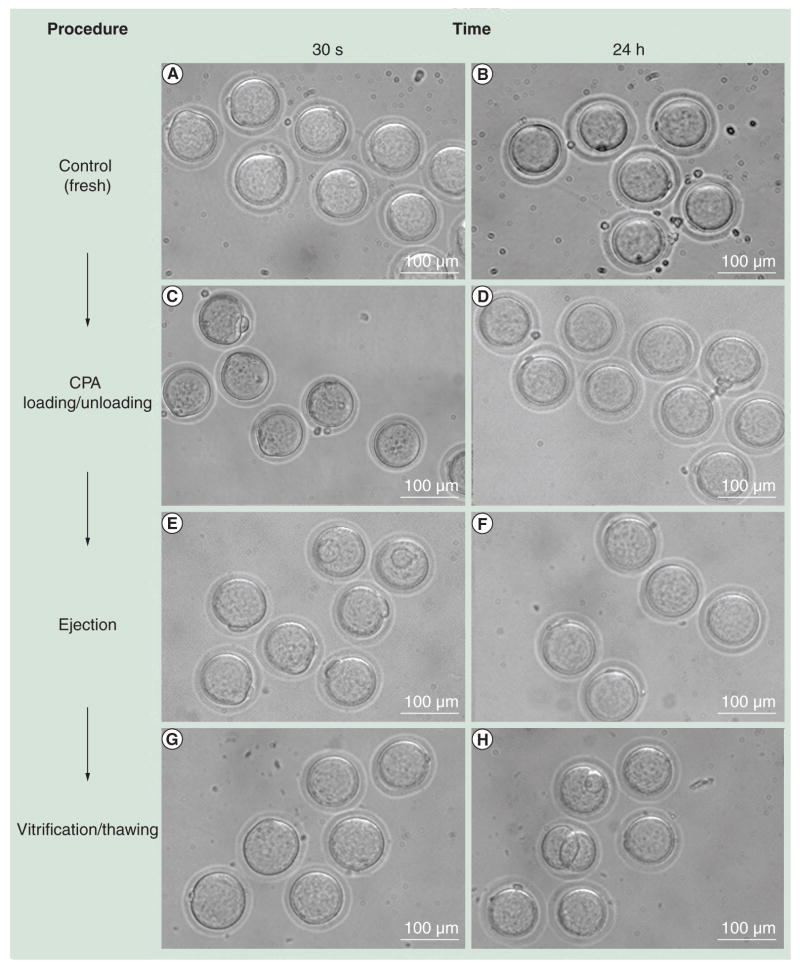
Figure 3. Morphological observations of oocytes at each procedure step.
Surviving oocytes from each procedure showed no difference compared with the controls in morphology. (A & B) Fresh oocytes (control); (C & D) oocytes recovered after CPA loading and unloading; (E & F) oocytes after ejection with CPA; and (G & H) oocytes after vitrification and thawing steps. Images were taken (A, C, E & G) 30 min and (B, D, F & H) 24 h after each procedure.
CPA: Cryoprotectant agent.

