
Fig. 3.
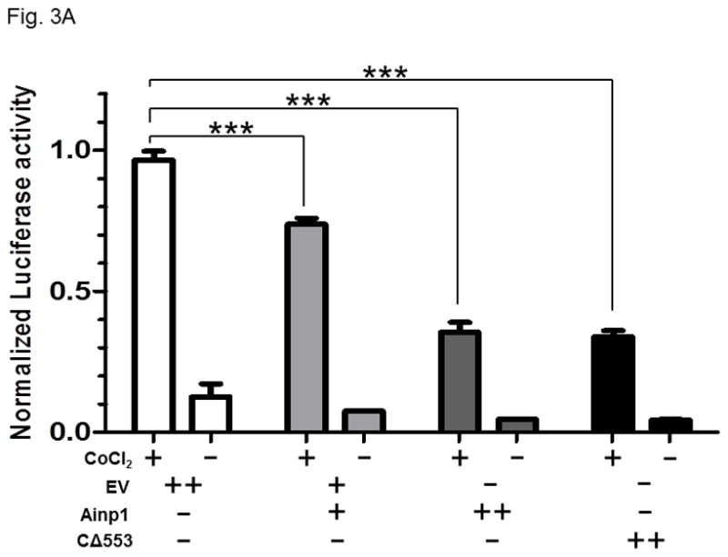
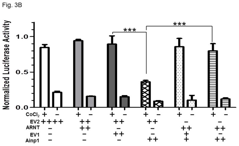
Ainp1 suppressed the CoCl2-activated, HRE-driven luciferase activity in Hep3B cells. A, cells (± 100 μM CoCl2) were transfected with the same amount of plasmid DNA (EV, pCMV- Tag4A; Ainp1, pCMV-Ainp1; C 553, pCMV-C 553). Amount of plasmid DNA used: +, 0.3 μg; ++, 0.6 μg; 150 ng of pGL3-Epo reporter luciferase plasmid; 50 ng of pCH110 plasmid. This experiment was repeated once with similar results. B, cells (± 100 μM CoCl2) were transfected with the same amount of plasmid DNA (EV1, pCMV-Tag4A; EV2, pSport; Ainp1, pCMV-Tag4-Ainp1; ARNT, pSport-ARNT). Amount of plasmid DNA used: +, 0.2 μg; ++, 0.4 μg; ++++, 0.8 μg; 75 ng of pGL3-Epo reporter luciferase plasmid; 25 ng of pCH110 plasmid. This experiment was repeated once with similar results. Luciferase activity was normalized by the β-galactosidase activity. Error bars indicate the variations of the means (mean ± SD, n = 3). ***p < 0.001.