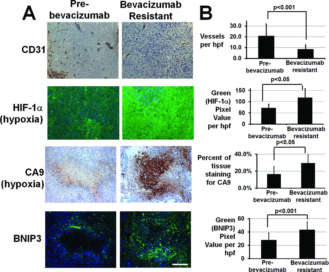
Figure 1. GBMs progressing during anti-angiogenic therapy exhibit decreased vessel density, increased hypoxia, and increased BNIP3 staining compared to pre-treatment GBMs from the same patients.
(A) Representative immunostaining for endothelial marker CD31 (upper row), hypoxia markers HIF-1α (second row; blue=Hoescht nuclear staining; green=HIF-1α), and CA9 (third row), and autophagy-mediating BNIP3 (lower row; blue=Hoescht nuclear staining; green=BNIP3) in this glioblastoma resistant to bevacizumab (right) compared to paired specimens from the same patient before treatment (left). CA9 and BNIP3 stainings are from adjacent sections. (B) Vessel density decreased (P<0.001), hypoxia marker HIF-1α staining increased (P<0.05), hypoxia marker CA9 staining increased (P<0.05), and BNIP3 immunostaining increased (P<0.001) in 6 GBMs after bevacizumab resistance compared to paired pre-treatment specimens. 40x magnification, scale bar=200 µm.

