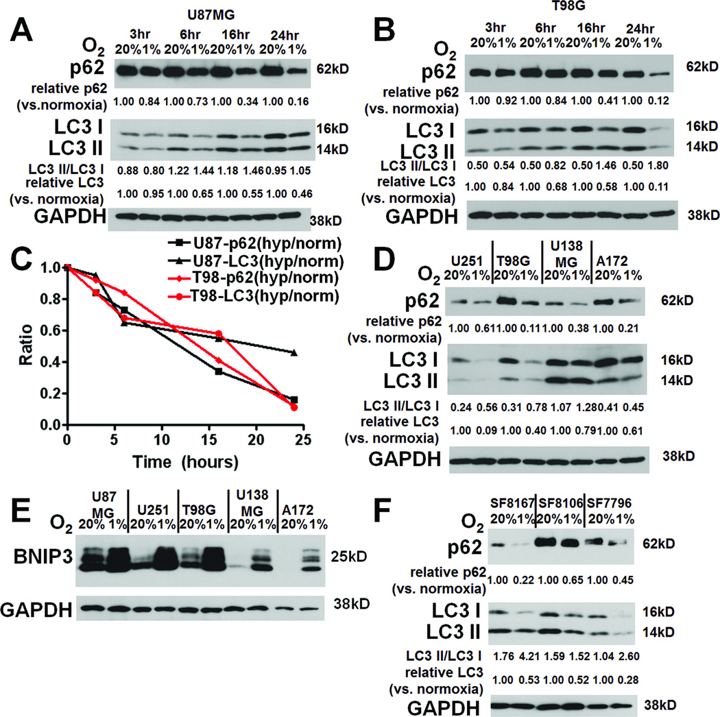
Figure 2. Hypoxia causes autophagy-associated protein changes in GBM cells.
Culturing U87MG (A) and T98G (B) GBM cells in hypoxia for up to 24 hours increased, relative to normoxia degradation of p62 and total LC3 (C), with T98G cells also showing increased conversion of LC3-I to LC3-II in hypoxia (B). Similar findings in 3 other glioma cell lines (U251, U138, A172, and G55) are shown at 24 hours (D). All cell lines also showed hypoxia-induced increased expression of autophagy-mediating BNIP3 (E). (F) Culturing primary glioma cells SF8167, SF8106, SF7796, and SF8244 led to the same hypoxia-induced autophagy-associated changes (p62 degradation, LC3-I to LC3-II conversion, and degradation of total LC3) after 24 hours.

