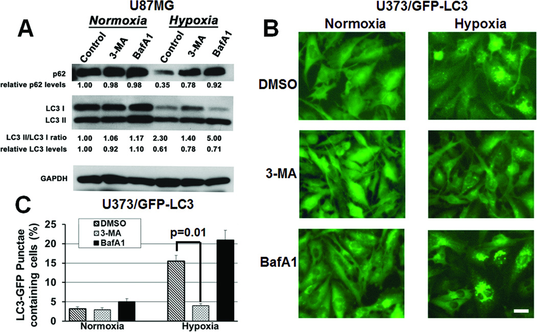
Figure 3. Autophagy inhibitors block the hypoxia-induced expression of autophagy mediators in glioma cells.
(A) After 24 hours, 3- MA (1 mM) and BafA1 (1 nM) blocked hypoxia-induced p62 degradation in cultured U87MG cells. 3-MA inhibited conversion of LC3-I to LC3-II, while BafA1 increased LC3-I to LC3-II conversion. (B) After 24 hours of hypoxia, U373/GFP-LC3 cells exhibited more punctate green fluorescent staining, consistent with autophagy, which decreased after 3-MA treatment, but remained high after BafA1 treatment. 40x magnification, scale bar=200 µm. (C) 3-MA reduced the percent of cells with over 10 punctate green fluorescent dots (P=0.01).

