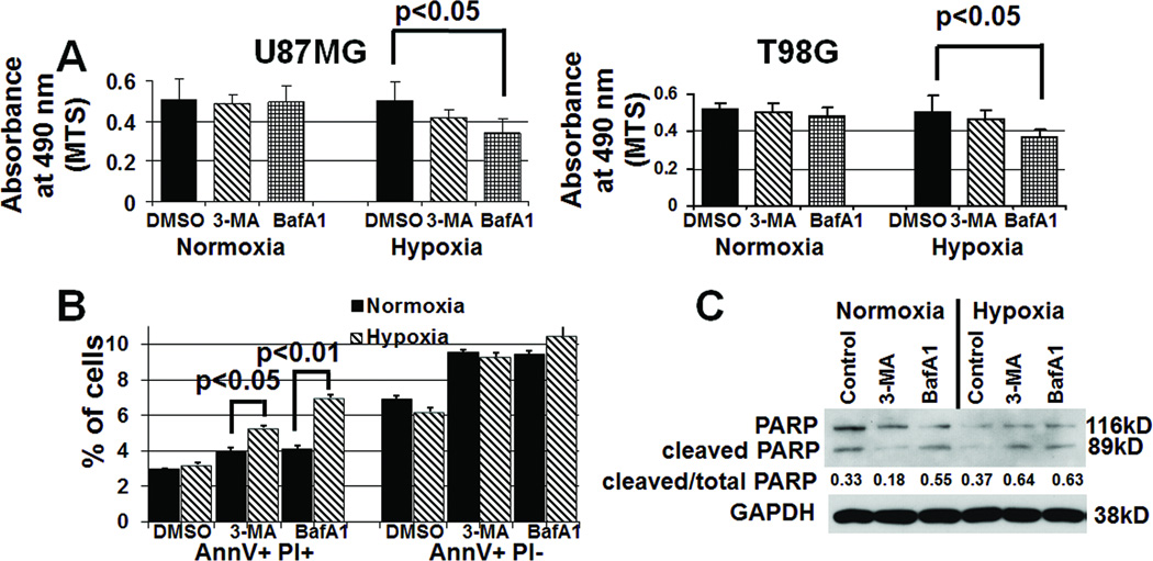
Figure 4. Inhibiting hypoxia-induced autophagy increases cell death.
(A) U87MG and T98G cells in hypoxia for 48 hours exhibited decreased cell numbers, as assessed by absorbance at 490 nm (reflecting number of cells) minus background measured in the MTS assay, when treated with 3-MA (1 mM) or bafilomycin A1 (BafA1; 1 nM) (P<0.05). (B) U87MG cells in hypoxia for 24 hours exhibited an increased percent of AnnexinV+PI+ cells (permeabilized near death cells, leftmost bars) in the presence of 3-MA (P<0.05) or BafA1 (P<0.01) relative to the presence of these inhibitors in normoxia, whilethese inhibitors did not change the percent of AnnexinV+PI− cells (early apoptosis, rightmost bars) in hypoxia (P>0.05). (C) Western blotting revealed increased cleavage of PARP, indicating apoptosis, in hypoxic cells treated with 3-MA or BafA1 for 24 hours, suggesting that autophagy inhibitors were promoting apoptosis.

