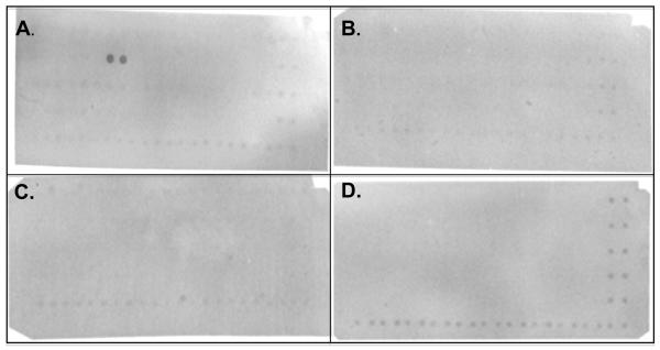
FIGURE 4.
Probing of SH3 domain array with the G9 monobody. Four membranes (purchased from Panomics) were incubated with His6-tagged SUMO fusion of the G9 monobody, washed, incubated with anti-His6-tag antibody conjugated to HRP, followed by detection with Enhanced Chemiluminescence-Plus (ECL-Plus). Panels A-D correspond to Panomics Arrays I-IV, respectively. Of the 150 SH3 domains tested, only the Fyn SH3 domain (duplicate, adjacent spots in panel A) bound to the G9 monobody. A His6-tagged ligand has been spotted in duplicate along the right side and the bottom of each membrane; these positive control spots are intended for alignment.

