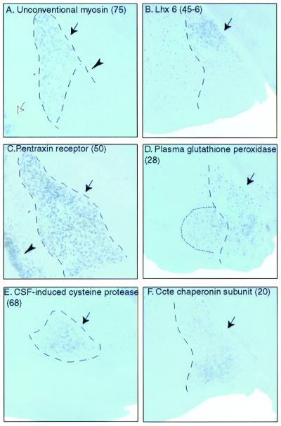
Figure 3.
Expression of amygdala-enriched genes in different amygdaloid subnuclei. (A) Probe 75 (unconventional type myosin, TIGR identifier TC37197). Note the sharp discontinuity in expression levels between the lateral (arrow), and basolateral (arrowhead) nuclei. Staining was also observed in cortical layers 2/3 (white arrow). No staining was detected in the cerebellum, olfactory bulb, or PAG (not shown). (B) Probe 45–6 (Lhx6, GenBank accession no. AB031040). Lhx 6 hybridized to many scattered cells in the forebrain and was particularly concentrated in the dorsal aspect of the medial amygdala (arrow); the cerebellum was unlabeled (not shown). Lhx 6 was not represented on the microarray, but was analyzed because of its coexpression with Lhx7 (28, 29), which also was enriched in the amygdala (not shown). (C) Probe 50 (neuronal pentraxin receptor, TIGR identifier TC18750). The expression domain matches the boundaries of the lateral and basolateral amygdala (arrow). Staining also was observed throughout cortex (arrowhead) and in hippocampus (not shown). No signal was detected in the cerebellum or PAG. The olfactory bulb had weak staining (not shown). (D) Probe 28 (plasma glutathione peroxidase, TIGR identifier TC31122). Intense labeling is apparent in the medial amygdala (arrow), hypothalamus, and PAG (not shown). Note also signal in a contiguous subregion of the basomedial amygdala (dotted line). Two other genes also showed expression in this same region (not shown). (E) Probe 68 [cerebrospinal fluid (CSF)-induced cysteine protease, TIGR identifier TC30215]. Hybridization in the basomedial amygdala (arrow) was detectable. Staining also was observed in the hippocampus but was absent in the remaining regions of study (not shown). (F) Probe 20 (Ccte chaperonin ɛ subunit, TIGR identifier TC30886). Signal was detected in the medial amygdala (arrow) and in the lateral, basolateral, and basomedial complexes (not shown). No staining was detected in the other four regions of study (not shown). Probe numbers are in parentheses.

