Figure 4.
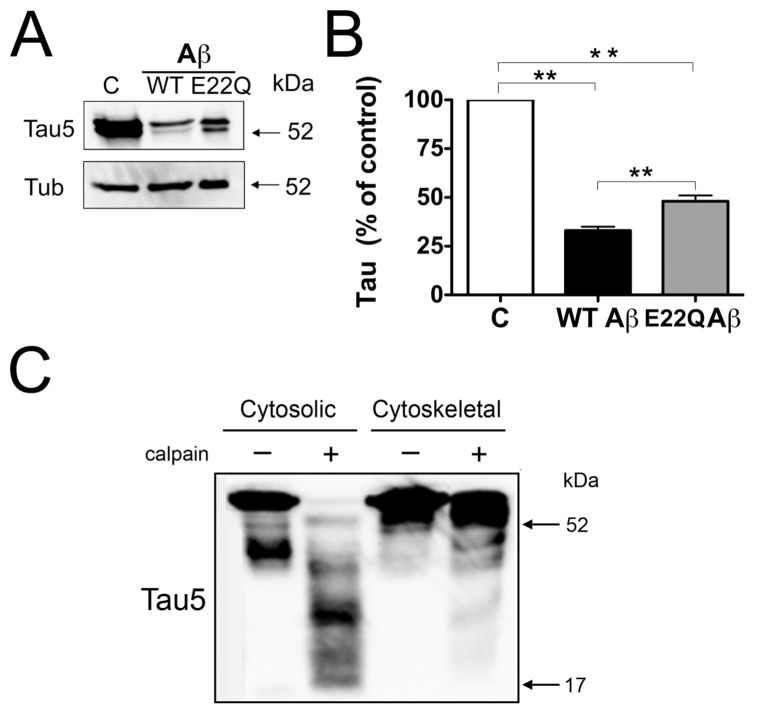
Decreased susceptibility of cytoskeletal-bound tau to calpain cleavage. (A) Western blot analysis of cytoskeletal fractions prepared from 21 d in culture hippocampal neurons incubated in the absence (C in panels A and B) or in the presence of WT Aβ or Dutch (E22Q) Aβ. The membranes were reacted with tau and tubulin antibodies. (B) Graph showing the results of the quantitative analysis of immunoreactive bands. Values are expressed as a percentage of the tau/tubulin ratio of untreated controls, considering the values obtained in these neurons as 100%. Each number represents the mean ± SEM from five different experiments. **Differs from control, P < 0.01. (C) Western blot analysis of cytosolic and cytoskeletal fractions obtained from untreated hippocampal neurons and then incubated in the presence (+) or absence (–) of active calpain. Membranes were reacted with a tau antibody (clone tau 5) and then stripped and reprobed with a tubulin antibody as a loading control. Note that microtubule binding decreased the susceptibility of tau to calpain cleavage.

