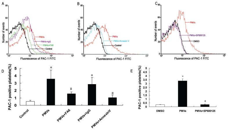
Figure 4.
CD36-, PS- and JNK-dependent platelet activation by PMVs. Platelets were incubated with 2 μg/mL FA6 (A,D), 2 μg/mL mouse IgG or 20 μmol/L SP600125 (C,E) before incubation with PMVs (30 μg/mL) and analyzed by flow cytometry with FITC-labeled PAC-1. In some studies, PMVs were first incubated with 20 μg/mL annexin V to mask PS (B,D). (A–C) Histograms represent MFI and graphs; (D–E) show percentage of PAC-1–positive platelets. Data are means ± SE from at least three separate experiments. *P < 0.05 compared with control; #P < 0.05 compared with PMV treatment.

