Fig 8.
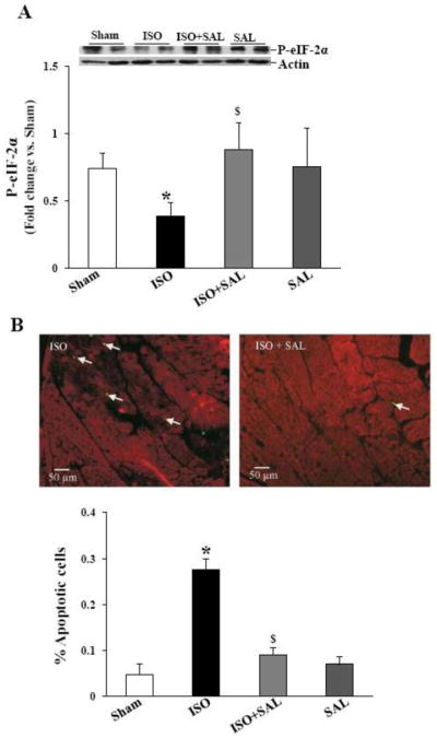
A. Salubrinal maintains eIF-2α phosphorylation in vivo. Mice were infused with ISO in the presence or absence of SAL for 7 days. LV lysates were analyzed by western blot using anti-phospho-specific eIF-2α antibodies. *p<0.05 vs. Sham; $p<0.05 vs. ISO; n=6–7. B. Salubrinal inhibits β-AR-stimulated apoptosis. Paraffin-embedded sections were used for TUNEL-staining assay. Upper panel demonstrates TUNEL-stained images obtained from ISO and ISO+SAL hearts. Yellow-green fluorescence represents apoptotic cells, while red fluorescence indicates α-sarcomeric actin staining (specific for myocytes). The lower panel demonstrates quantitative analysis of myocytes apoptosis with or without ISO-treatment; *p<0.05 vs sham; $p<0.05 vs. ISO; n=3–5.

