Figure 6.
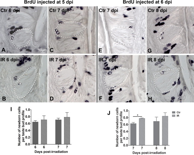
The influx of new cells into taste buds resumes and is accelerated at 6 dpi. A–D, BrdU was injected at 5 dpi, and newborn cells inside taste buds quantified at 6 dpi (A, B) and 7 dpi (C, D) in irradiated and sham-irradiated groups. E–H, BrdU was injected at 6 dpi, and newborn cells assessed at 7 dpi (E, F) and 8 dpi (G, H) irradiated (IR) and control (Ctr) CVP taste buds. Dashed lines indicate borders of taste buds. White asterisks mark newborn cells inside taste buds. The influx of cells into taste buds was significantly accelerated at 7 dpi and assayed at 24 h following BrdU injection at 6 dpi (J), but not at any other time points (I, J). t test, n = 3 mice, and 60–183 taste buds tallied per time point and condition, *p < 0.05.

