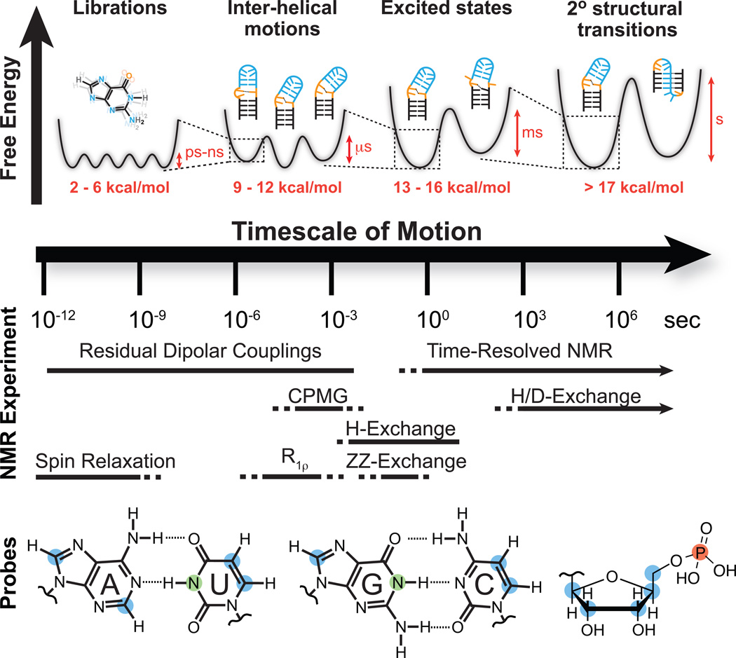
Figure 1.
NMR techniques and site-specific probes for characterizing motional modes that carry RNA structure along various regions of the dynamic structure landscape. Dynamic Structure Landscape: Transition free energies (red, kcal/mol) corresponding to typical timescales of interconversion were estimated using transition state theory at 25 °C. NMR Experiments: Solid lines indicate the timescales at which each NMR experiment is optimally suited for, while the dotted lines indicate timescales that are difficult to probe. Probes: Nuclei most commonly used for RNA dynamics measurements: protonated carbons (blue), imino protons (green), backbone phosphorus (red).

