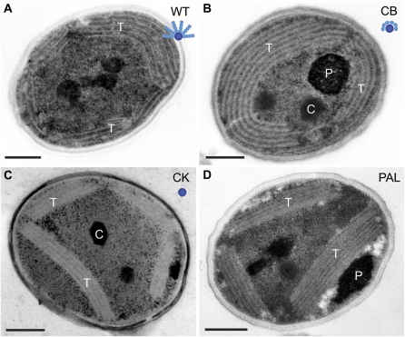
Figure 2.
Thin-section electron micrographs of Synechocystis 6803 wild type and phycobilisome antenna mutants. A, Wild-type (WT) strain with intact phycobilisomes. B, CB mutant with one PC hexamer per rod. C, CK mutant that contains only the APC core. D, PAL mutant that lacks the assembled phycobilisomes. Labeled are thylakoid membranes (T), polyphosphate bodies (P), and carboxysomes (C). A cartoon model of the phycobilisome structure in each strain is shown as an inset. The APC core is represented by the central blue circle, and the PC discs are shown as teal ellipsoids. Bars = 250 nm. [See online article for color version of this figure.]

