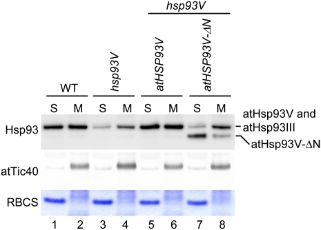
Figure 6.
atHsp93V-ΔN has reduced membrane association in vivo. Chloroplasts isolated from 14-d-old plate-grown plants of the indicated genotypes were lysed hypotonically and separated into soluble (S) and membrane (M) fractions by centrifugation. The proteins in the soluble fraction were precipitated by TCA and dissolved with the protein extraction buffer. The membrane fractions were resuspended with the same volume of protein extraction buffer. Protein concentrations were then determined for all samples. Eight micrograms of proteins was loaded in all the soluble-fraction lanes. For each membrane fraction, the same volume as its corresponding soluble fraction was loaded so that each pair of membrane and soluble fractions shown were derived from the same amount of chloroplasts. Samples were analyzed by SDS-PAGE. The top half of the gel was analyzed by immunoblotting with antibodies against Hsp93 and atTic40. The bottom half of the gel was stained with Coomassie blue to reveal the location of RBCS. WT, Wild type. [See online article for color version of this figure.]

